
Each of eyes contains an area that has no photoreceptors because it is occupied by the optic nerve. This area is known blind spot. These areas are on opposite sides of visual field. Following exercise is isolate the blind spot.
Instructions: Close left eye and fix right eye on the cross. Place eyes about 12 inches (30 cm) away from the monitor (distance may vary depending on the screen resolution) and notice the dot disappears. .

Note that the dot is replaced, not by a black region, but rather blank white space. This is because the brain simply "fills in" the most probable stimulus (in this case, a uniform white area) where there is none.
The following examples demonstrate the "filling-in" phenomenon in greater detail. Apply the same instructions as given above and notice the red markings each time are replaced by the most probable pattern that your brain is able to perceive.


Try and count the dots in the diagram below. Despite a static image, eyes will make it dynamic attempting to "fill-in" the white circle intersections with the black of the background. Quite an amazing effect!
Instructions: Simply stare at the white circles and notice the intermittent blinking effect.

Colors often appear brighter and more vibrant when they are bordered by frames. Black lines are commonly used to enhance colors in applications like stained glass. This tactic creates a certain effect, as shown below, and prevents color clashing. Notice that the drawing on the left colors appear significantly brighter and pure.


This "adaptation mechanism" allows eyes to recover from an oversensitivity to a particular stimuli. "Chromatic adaptation" occurs when eyes adjust to certain color stimuli. For example, when entered in a movie theater on a sunny afternoon, room appeared dark but as visual system adjusted to dim lights we were able to see better again.
Follow the instructions below and see how the visual system responds to a color overload.
Instructions: Fix eyes on the black spot in between the uniform cyan and yellow areas for about 30 seconds. Then look down and shift your gaze to the black spot in the 2nd image. Note that the image of the seaplane appears approximately uniform after this adaptation.

Almost 10% of human males experience color vision deficiency (compared with 0.4% of females). The most common form of these abnormalities is characterized by an inability to distinguish between red and green hues.
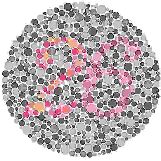
Instructions: The following images are part of The Series of Plates Designed as a Test for Colour-Deficiency by Shinobu Ishihara M.D. which is the accepted standardized color blindness test. They are specially adjusted to isolate the exact deficiency experienced by the viewer. What do you see in the plates?
This is a test plate in which everyone should see a "12".

This plate is designed to separate the type of color defectives and the level to which they are observed. Most will see the number "26" clearly while some will only see a "2" or a "6" or no numerals at all.

Can you trace a line from one "X" to the other? Someone with normal color vision will trace a orange/brown purple line and those with a slight deficiency will follow a different path.

Simultaneous Contrast (colors taking on characteristics of their complement) is occasionally take precedence over by the Spreading Effect. This occurs time and again when there is a difference in the "spatial frequency" of objects on a background. After analyzing the diagram below, see how this tactic can be applied to the design of tapestries in order to preserve certain color sensations.
Instructions: Look at the two sets of gray patches occupying the same area space on the light red background. Notice that the patches on the left appear slightly greenish. On the right, the gray patches definitely tend towards red. Simultaneous contrast is taking place on the larger blocks of the neutral gray. Whereas, the thinner strips of gray exhibit "spreading."

Dithering is a color reproduction technique in which dots or pixels are arranged in such a way that allows to perceive more colors than are actually used. This method of "creating" a large color palette with a limited set of colors is often used in computer images, television and the printing industry.
The graphic illustrations below show how a full color image can been approximated by using only a few colors. As magnification is increased, see how each color seen is broken down to a combination of the primary colors used in the dither pattern.
 |
 |
| Original Image | Grey Image |
 |
 |
| Dithered Image at 100% scale | 200% scale |
 |
 |
Understanding how the eyeball interprets color is essential for creating color palettes in certain media. Color printers, which typically use 3-4 colors (sometimes more), and televisions, which use red, green and blue only, are a couple examples of devices that use dots or pixels to display color.
Instructions: The following series of images begins with a collection of dots that are clearly green and have significant amounts of space in between them. Reducing the original image, we notice that the colored dots seem to blend together and a coherent color appears solid in the final image. Using similar methods, it is possible to mix colors by simply altering dot formations and limiting white space. Both RGB (television, computer monitors, film projection) and CMYK (4-color process) use this technique.


Although there are no actual triangles that appear on your eyes' retinas, your brain will somehow interpret the following image as two overlapping triangles. Is this imagination? Are you losing your mind? No, the notched circles and angled lines merely suggest gaps in which objects should be. The brain does the rest by triggering a sort of pattern recognition phenomenon.

The nature of our visual system allows us to sometimes see "after-images" which appear once the original stimuli are removed. In the following demonstration, you will see that the colors in after-images are usually the opposite (complementary) colors of the original.
Instructions: Stare at the black spot in the center of the four colored squares for about 30 seconds. Then scroll down and move your gaze to the black spot in the uniform white area. Note the colors of the afterimages relative to the colors of the original stimuli. Did they appear different?

During the Optical Art (OpArt) Movement of the 1960s, artists would create all sort of puzzling effects with color. For instance, this "flashing squares" drawing seems to wobble and flash when you concentrate on one particular area of the image. How many squares can you see in this diagram? Can you feel the "motion" of the image?

How objects and colors appear is highly dependent on their context. The structural and spatial variables of a scene can influence appearance and perception. The following optical illusion demonstrates how we are sometimes fooled by our eyes.
Instructions: The diagram below features two circles with different surroundings. Would you believe that the two circles are identical?

Identical colors appear to shift when framed by different backgrounds or patterns. This is called "simultaneous contrast" and has a variety of affects on how we see things.
Instructions: The diagrams below feature two sets of identical red and green squares within a striping pattern. Do the colors on each side of the stripes appear different? In each case, the squares on the left side appear darker and the right side appears lighter. View the images from the side of your monitor to exaggerate this effect.

Viewing two colors at the same time influences both of their appearances. The following is an example of induction, a variation of simultaneous contrast.
Instructions: In the top half of the diagram below are two identical dark gray patches on a like background. The bottom half of the diagram shows the same dark gray patches on different backgrounds. You will see that they appear different because of their surroundings.
